What is a cell? The IMAT-level answer
A cell is the basic structural and functional unit of living organisms. Cells are enclosed by a plasma membrane and arise from existing cells. For studying cell biology, distinguish prokaryotes from eukaryotes, and a membrane from a wall. These are general organisational patterns: specialised cells can lose structures, so the absence of a nucleus alone does not prove that a cell is prokaryotic.
Cell biology connects structure with function. A useful starting point is learning which features distinguish cell types, and which apparent rules have exceptions.
Cell theory in one paragraph
Three claims, and the exam expects all three:
- All living organisms are made of one or more cells.
- The cell is the basic unit of structure and function in life.
- All cells arise from pre-existing cells.
The third point is the one that carries the most weight historically — it is what ended the idea of spontaneous generation. It is also the reason viruses sit outside cell theory: a virus has genetic material and a protein coat, but it cannot make proteins or produce energy on its own, and it does not divide. It replicates by hijacking a cell that does.
Prokaryote versus eukaryote
The table describes typical cellular organisation, not every specialised cell.
| Prokaryote | Eukaryote | |
|---|---|---|
| Nucleus | None — DNA lies in the nucleoid region | Nucleus with a double membrane in typical cells |
| DNA | Typically circular chromosomal DNA in a nucleoid | Linear chromosomes wound on histones |
| Membrane-bound organelles | None | Mitochondria, ER, Golgi, lysosomes… |
| Ribosomes | Present — 70S | Present — 80S in the cytoplasm |
| Typical size | 1–10 µm | 10–100 µm |
| Cell wall | Usually present (peptidoglycan in bacteria) | Plants, fungi yes; animals no |
Two traps live in that table.
Prokaryotes do have ribosomes. “No membrane-bound organelles” is often misremembered as “no organelles at all”. Ribosomes are not membrane-bound, and cells need them to synthesise proteins. Mature human red blood cells are a specialised exception: they have lost their nucleus and ribosomes and cannot make new proteins.
Mitochondria and chloroplasts have their own ribosomes. These differ from cytoplasmic ribosomes and support bacterial ancestry, alongside other evidence. Do not assume that every mitochondrial ribosome has the same sedimentation value.
Plant versus animal cells
Both are eukaryotic, so the differences are additions, not upgrades.
A typical mature plant cell has a cellulose cell wall and a large central vacuole that supports turgor. Photosynthetic cells contain chloroplasts; many root cells do not. An animal cell has centrioles and typically more prominent lysosomes.
The classic error: assuming plant cells have no mitochondria because they photosynthesise. They have both. Photosynthesis makes glucose; respiration releases the energy from it, and plants respire around the clock.
Another: treating the cell wall as “the plant version of the cell membrane”. Plant cells have both. The membrane controls what enters and leaves and is selectively permeable; the wall is a rigid outer support that is freely permeable to water and small solutes.
The organelles worth knowing properly
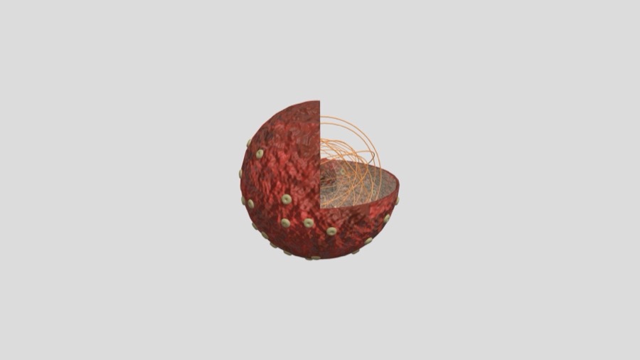
- Nucleus — holds the DNA; the nucleolus inside it assembles ribosomes.
Drag to rotate · scroll to zoom
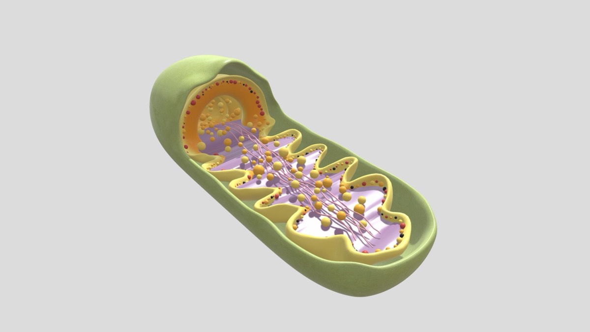
- Mitochondrion — aerobic respiration; double membrane, inner one folded into cristae to increase surface area; has its own DNA.
Drag to rotate · scroll to zoom
- Rough ER — studded with ribosomes; processes proteins destined for secretion or membranes.
- Smooth ER — lipid synthesis and detoxification; no ribosomes.
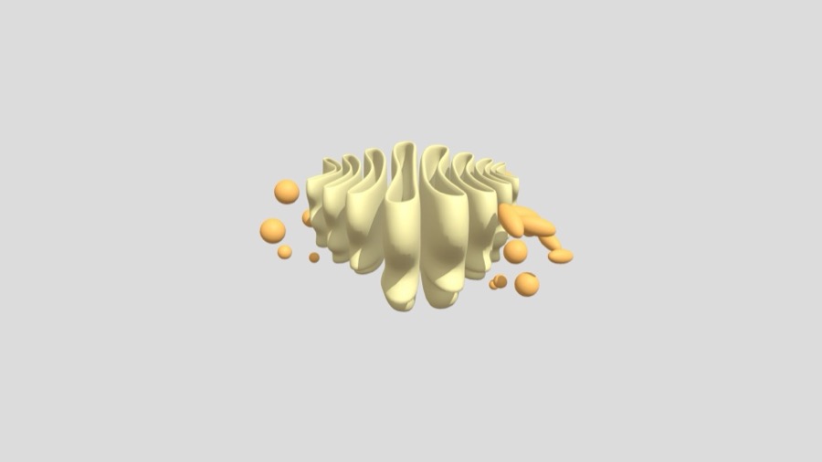
- Golgi apparatus — modifies, sorts, and packages what the rough ER sends.
Drag to rotate · scroll to zoom
- Lysosome — hydrolytic enzymes in an acidic interior; breaks down waste.
A useful application is to trace a secreted protein through the cell. Ribosome on the rough ER → ER lumen → vesicle → Golgi → vesicle → plasma membrane → outside. If you can order that pathway, you can answer a whole family of questions.
What the exam actually does with this
Recognition questions (“which organelle contains hydrolytic enzymes”) are the easy end. The harder pattern gives you a function and asks which structure must be present — or gives you an organism and asks what follows from it.
A cell with no nucleus, a wall and 70S ribosomes fits a prokaryotic pattern, but those features alone do not distinguish Bacteria from Archaea. A peptidoglycan wall is additional evidence for Bacteria. Likewise, a mature human red blood cell has no nucleus but belongs to a eukaryotic organism. Use several features together.
The five confusions that cost marks
- Prokaryotes have no ribosomes. (They have 70S ribosomes.)
- Plant cells have no mitochondria. (They do — they respire too.)
- Cell wall and cell membrane are the same thing. (Plants have both.)
- Viruses are very small cells. (They are not cells at all.)
- The nucleolus makes DNA. (It assembles ribosomes.)
Each of these is a belief, not a gap — which is why re-reading the chapter rarely fixes them. What fixes them is meeting the contrast head-on.
Sources
Or keep going in the iPhone app.
Found a mistake? Write to hello@imatlearn.com — corrections are made at the source.